
It is an interesting and intuitive function of a tree’s vascular system to have nourishment blindspots–areas where the vascular system dies back due to, essentially, having dead ends in their vascular system. They can form as a result of pruning, breakages, or pathogens. They’re often visible on the bottom-side of an old pruning cut of live tissue from a woody plant.
Blindspots don’t always form after pruning cuts, and they aren’t of major consequence to the plant either. The only comment against them is that with increased surface area, the pruning wound will take longer to compartmentalize. Sometimes this happens when the removed limb lacked a branch collar. But it isn’t as simple as the branch dying back to its internal attachment point. The integration of branch tissue with trunk tissue is complex.
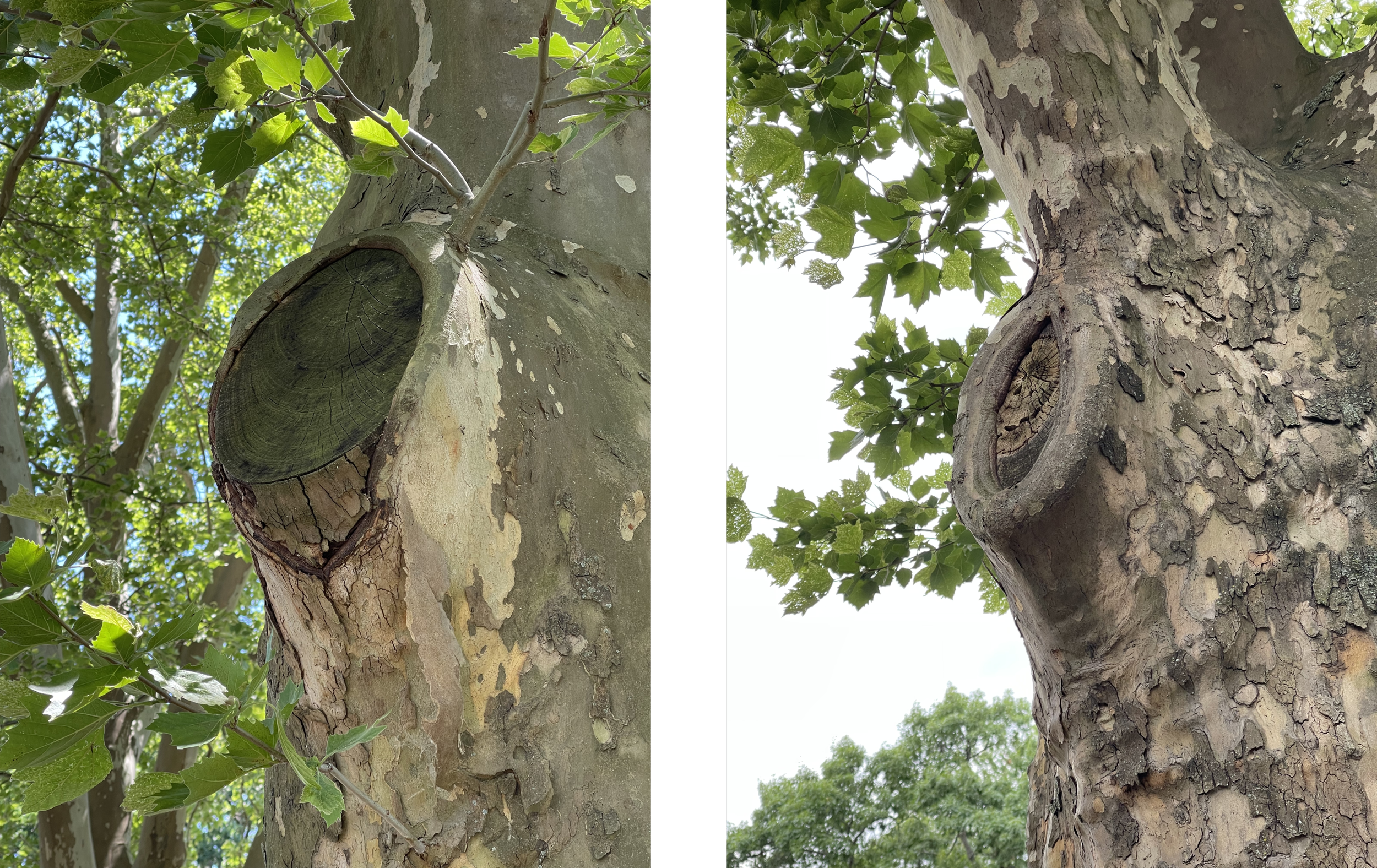
IMAGE 1: Left: wound with a nourishment blindspot its bottom margin. Right: An older wound that did not form a blindspot. Both Platanus x hispanica.
Both cuts in Image 1 were done properly by modern standards, evidenced by the cut surface being visible (pictured in yellow below). Both were living branches when they were cut. Why then, did the trunk tissue die back below the cut on the left, and not on the right?

IMAGE 2: The yellow surface is the where the cut was made. The red area is the nourishment blindspot.
The reason for blindspots (pictured in red above) can be explained by looking at the general flow of the vascular system, along with some of the basic features of the cells within it.
Vascular System Flow
The conducting parts of a tree’s vascular system are most active around the perimeter of the stems. Xylem and phloem, and a few other cell types and structures, are found here. Their primary functions are the movement of water and carbohydrates throughout the tree.
Flow
The vascular system is much more tortuous and complex than it is depicted below, but for the sake of this explanation we just need to see the general flow of things.

From left to right:
the left image represents the vascular system when the branch was intact, prior to being cut.
the middle image is the flow of the vascular system immediately following the cut.
the right represents how the vascular system’s flow has since diverged.
It is the divergence in the third image that forms the blindspot, which results when the vascular cells in the middle image have nowhere to conduct to: they dead end. The vascular system “dies back” to the closest point where the cells can properly reroute fluids around the blindspot.
I suspect there are many variables that influence the presence of a nourishment blindspot:
Temperature when the wound/cut is made
Shape & size of the wound/cut
General vigor of the tree
Species & cell characteristics
Presence/position of a branch bark ridge
Colonization of pathogens
Cells
There are many different cells and cell types in a woody plant’s vascular system. For simplicity’s sake, only xylem are examined here. First we’ll look at the individual xylem cell, and then zoom out a bit.

IMAGE 3: Diagram of the relevant characteristics of xylem cells
Xylem Cell
There are many organelles and features in xylem cells. Image 3 highlights three of the characteristics of the xylem cells that make the rerouting of fluids possible.
Lumen: A hollow channel running through the length of the cell, through which is the major pathway for water.
Pits: Small holes which connect the lumen of adjacent xylem cells together.
Perforation Plate: A grate-like structure that connects the ends of xylem lumen together. It helps prevent cavitation & embolism (or air) from spreading vertically, which would significantly disrupt continuity.
Zooming Out

IMAGE 4: LEFT: Sagittal view of a lattice of active xylem working together before a cut is made above them. RIGHT: View of the lattice after the cut, showing the yellowed cut area above the red blindspot area.
In xylem shown on the right of Image 4, the yellow region is the cut surface, and the red region is the nourishment blindspot, just like in Image 2. Notice the cells in the red region dead end. They have no continuous path to follow, and so embolize, causing the “dieback” to a point where the perforation plates, together with the pits, can maintain a cohesive water path through a tree.
It is partially due to the pits and perforation plates that the entire vertical column of xylem beneath the cut isn’t embolized (also due to some other cool cell capabilities). Without these tiny features, each wound a tree was dealt would result in significant hydraulic failure.
More Thoughts
If you are someone who works with trees consistently, you’ve recognized different species have different characteristics when it comes to their responses to being pruned.
One contributing factor to that variability is cell morphology; the characteristics of cells. Some species have vascular cells with wider lumens, some have differently shaped perforation plates, and/or different number of pits. Each of those alters how a tree handles cavitation, and thus pruning.
I’m not really sure if this information has much in-field application. I think it’s just cool to think about trees. And I hope you find it cool too.